


Tumor Treating Fields (TTFields) are a treatment method for different tumors e.g. glioblastoma, a highly malignant brain tumor [4]. TTFields are harmonic CW electromagnetic (EM) fields in the frequency range between 100 kHz to 500 kHz which are enabled through electrode arrays and irradiate the afflicted (brain-) region (see Figure 1). Tuned to proper field amplitude, frequency and polarization the electric fields inhibit the propagation of tumor cells which are mitotically active . Compared to other tumor treatment methods, a huge advantage of TTFields is that they have almost no effect on healthy cells and the whole organism, resulting in no side effects for the afflicted. Although several studies show the efficacy of TTFields concerning overall survival and progression free survival [3, 4], the discussion of the mechanisms of action of TTFields is not finally settled. A fundamental understanding of the mechanisms of action from TTFields on cells would possibly allow the development of enhanced applications for tumor treatment. At the Institute of Microwave and Wireless Systems these mechanisms of action are systematically investigated. In order to find the physical interaction of TTFields with cells numerical electromagnetic simulation software is applied and electric equivalent circuit models are developed. In order to evaluate theoretical results, in-vivo and in-vitro experiments are conducted by our research cooperation partners. The according TTFields application setups are designed and implemented at the Institute of Microwaves and Wireless Systems.
Numerical EM Simulations of Cells under TTFields Treatment
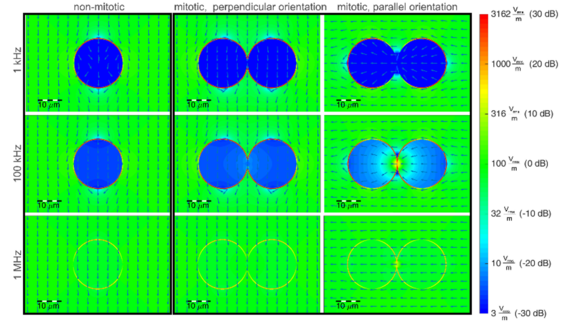


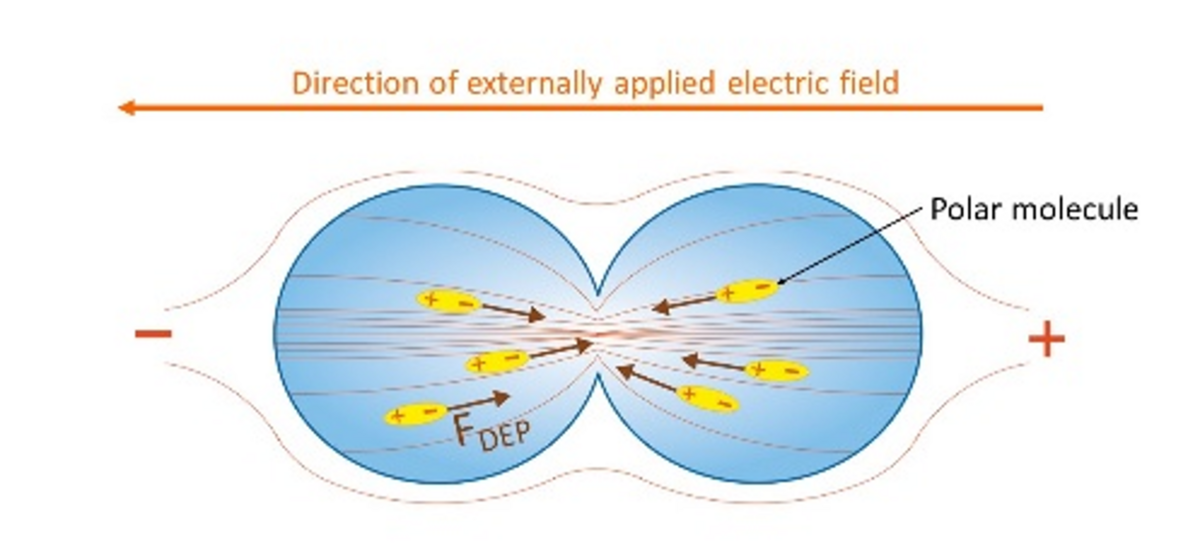
Figure 2 shows the numerical EM simulation of a single cell (non-mitotic) and of a dividing cell (mitotic) under TTFields treatment. Here, a simplified cell model is used. The shape of the cell model is spherical and it consists of a cell membrane (capacitance C = 29 pF) and a cytoplasm (εr = 80, σ = 1.3 S/m). The capacitive property of the cell membrane shields the interior of the cell from electric fields in low frequency ranges. As the frequency increases, the cell becomes permeable for the electric field. In the case of a mitotic cell, there is a relatively high electric field gradient at the cytokinetic cleavage furrow (CCF) when the electric field is polarized parallel to the mitotic axis. This gradient in electric field is assumed to cause dielectrophoretic forces (s. Figure 3) on cellular compartments which are polarizable or polar, as tubulin dimers.


Furthermore, in the case of the electric field oriented parallel to the mitotic axis, there is also a relatively high field magnitude in the CCF which results in a relatively high specific absorption rate (SAR), probably causing heating within this region. However, the cell model used here is a vast simplification of a real cell structure. By considering other cell compartments as well, further possible mechanisms of action can be worked out and evaluated in terms of significance in the frame of TTFields and cancer treatment.
Equivalent Electric Circuit Model of Cells in Cytokinesis
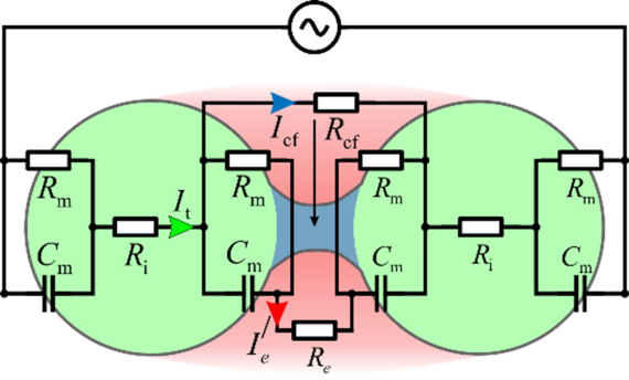
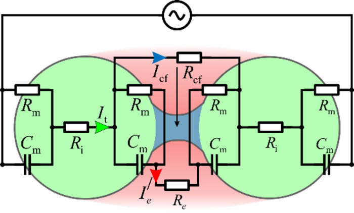

Figure 4 shows an equivalent electric circuit model of a dividing cell. The two daughter cells in this model consist of a cell membrane (Cm and Rm) and a cytoplasm (Ri). They are connected by the cleavage furrow (Rcf) and located in an extracellular medium (Re). The lumped element parameters within this model depend on the electric properties of the respective cell compartments and their size. Thus, this model can be extended to several different cell types and provides the possibility to relate the electric behavior of the cell to the considered compartments.
Setup for in-vitro experiments
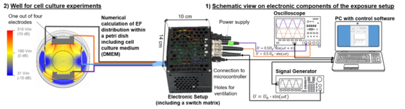
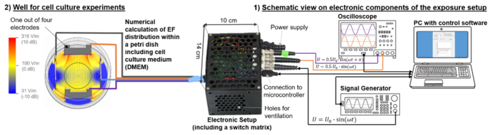

Figure 5 shows a schematic view on the experimental setup for in-vitro experiments with cells. Before designing and implementing electronics for this setup, the desired field distribution within the cell culture well has to be defined. In this version (s. Figure 5, left side numerical simulation [2]), two different polarization directions of the electric field vector are enabled by four electrodes. Furthermore, the cell cultivation area is located in a determined area (green circle within cell culture well) in order to irradiate cells under test with an almost homogeneous electric field (with magnitude ERMS = 100 V/m). The design and implementation of the electronics as well as the programming of the graphical user interface for controlling complement the whole setup.
Our cooperation partners
In cooperation with the Institute of biophysics of Leibniz University of Hannover and the neurologic clinic of the Hanover Medical School various different in-vitro and in-vivo experiments are conducted in the frame of TTFields research. The three cooperation partners complement each other with their expertise on the different major fields important for TTFields research.
Responsible Research Assistants


30167 Hannover
References
[1] Berkelmann, L., Bader, A., Meshksar, S., Dierks, A., Hatipoglu Majernik, G., Krauss, J. K., Schwabe, K., Manteuffel, D., and Ngezahayo, A. 2019. Tumour-treating fields (TTFields): Investigations on the mechanism of action by electromagnetic exposure of cells in telophase/cytokinesis. Scientific reports 9, 1, 7362.
[2] Berkelmann, L., Bader, A., Meshksar, S., Ngezahayo, A., and Manteuffel, D. In Vitro Exposure System for Tumor Treating Fields. IEEE International Engineering in Medicine and Biology Conference 2019.
[3] Weinberg, U. Novocure’s Tumor Treating Fields: Innovative brain cancer therapy with survival and safety benefits. https://www.nature.com/articles/d42473-018-00156-3. Accessed 16 May 2019.
[4] Zhu, P. and Zhu, J.-J. 2017. Tumor treating fields: a novel and effective therapy for glioblastoma: mechanism, efficacy, safety and future perspectives. Chinese clinical oncology 6, 4, 41.


